N.B. Nair
Berlin (ISJ): Study of molecules requires mapping of electron dynamics, whether it is in chemical reactions, biological processes or many high-tech applications. A team of scientists at Max Planck Institute of Solid State Research in Stuttgart, Germany has developed the fastest microscope that can map electron dynamics in space and time.
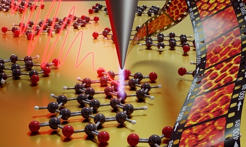
Currently, microscopy methods only provide sharp images either spatially or temporally. But a team lead by Dr Manish Garg combined established techniques of tunnel microscopy and laser spectroscopy to overcome these shortcomings. Tunnel microscope is used for imaging surfaces at the atomic level. Their innovation can visualise the movement of electrons in individual molecules. Existing methods could not image fast electrons directly, but have always had to resort to techniques only to reconstruct the behaviour of electrons.
The microscope developed at Max Planck Institute would also help understanding the biological processes such as plant photosynthesis, for development of solar cells or novel electronic components.
Although modern microscopy methods offer almost unlimited possibilities, they are always associated with certain compromises. For example, scanning tunnelling microscopy with a resolution of one tenth of a picometer (a picometer is one trillionth of a meter) allows extremely sharp images of individual atoms to be taken. But it is slow and cannot capture the electron dynamics in a material. Optical methods with ultrafast laser pulses, on the other hand, can detect electron movements in the attosecond range, but only provide spatially thinned out images – far beyond the atomic resolution that is possible with scanning tunnelling microscopes. One attosecond corresponds to one billionth of a billionth of a second. The typical electron dynamics and laser pulses are in the range of a few hundred attoseconds.
“We have been working for several years to combine these two techniques in such a way that each can play to its strengths without bringing in its weaknesses," said Dr Manish Garg, head of a research group at the Max Planck Institute for Solid State Research.
The researchers had to combine the tried and tested scanning tunnelling microscopy with state-of-the-art laser technology. In a scanning tunnelling microscope, a wafer-thin, atomically thin tip travels just above a conductive surface. Thanks to the quantum physical tunnel effect, electrons can flow between the surface and the microscope tip, even if there is no direct contact. For example, a molecule on a surface can be gradually roasted atom by atom.
The new microscopy technology now uses laser pulses to modulate the tunnel current by specifically excitation of the electrons in the material. "This has to be done extremely quickly, otherwise thermal effects will come into play and make the measurements impossible," explained Alberto Martin-Jimenez, a scientist involved in the experiments. The necessary ultrafast laser pulses in the attosecond range are not available off the shelf. But thanks to the rapid development of laser technology in recent years, the researchers have now succeeded in generating exactly the right pulses. Two years ago, Garg and Klaus Kern, Director at the Max Planck Institute demonstrated the function of such an atomic quantum microscope for the first time.
"For the first time, we were able to directly image the dynamics of electrons in molecules as they jump from one orbital to another," said Garg. "This basic technology provides completely new possibilities for directly observing quantum mechanical processes such as charge transfer in individual molecules and thus better understanding them."
The possible areas of application for such a quantum microscope are still hardly foreseeable. Especially in charge transfer processes, as they play a decisive role in many biophysical reactions as well as in solar cells and transistors, it could provide decisive new insights.
Source: Dr Manish Garg, Max Planck Institute
Image courtesy: Dr Manish Garg


